Physician's Notebooks 9 - http://physiciansnotebook.blogspot.com - See Homepage
6a: The Lobes of the Brain -Update 04 Septr 2021
The following descending table has main headings that you may search & find or scroll to:
A view of a Brain's Surface
Specialization of functions
Keep in Mind: 3 more chapters on cerebral cortex follow this one
The surface-wrinkled part of the human brain is the center of interest in neurology, psychology, psychiatry. It is the source of the almost impossible to describe but constantly experienced thing called consciousness. It stores our memories, it directs our attention to needles in haystacks, it causes us pleasures and pains, it can make us want to enjoy life or to commit suicide.
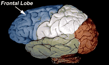
A view of a Brain's Surface: Above you see the Brain from its body's left side, facing to your left. This view shows the brain's left hemisphere, lateral and superior surface. Note the 4 flashing names of the lobes. In front and upper (in blue) is frontal lobe - the prefrontal & orbitofrontal part overlying the eye orbit is the source of executive action and, on its inside, the medial part, contains the working memory. The more rear part is the source of motor control. The frontal lobe border in its rear with the parietal lobe (the lobe in white) is the fissure of Rolando. (Not labeled above; it separates the frontal blue from the more rear white surface) The motor area is the gyrus (the rounded wrinkled ridge-strip) in front of the fissure.
The parietal lobe gives one's consciousness an awareness of its space relations and, working with the occipital lobe, it helps interpret our visual fields. And it brings feelings of touch and pressure and position into consciousness. It is the so called "where" area because it computes where things we see are located. It also directs our attention to needles in haystacks.
The occipital lobe (in red), registers at the first level of visual integration of the visual images that fall on the eyes' retinas.
The temporal lobe (in gray) on the side and below is the source of emotions, hearing and, and on its inner side (limbic lobe) of long-term memory and, relating to that, of heard speech. Its inner, lower part - the so called "who" area - identifies who we see - like recognizing individual faces.
The cerebral cortex shows localization of function but it also functions with other parts of brain in a distributive way. For example, memory is distributed in many parts of the brain - working memory in pre frontal lobe, declarative long-term memory in median temporal lobe, motor-measure memory in cerebellum. And in vision, the various parts of vision - color, illumination, shape, movement - are distributed in many parts of the parietal and occipital lobes and then brought together in a single visual instant in our consciousness. The point here is that the brain has multi-modal wiring systems that feature both labeled lines (separation as parallel passages of various sensations like vision, hearing, smell from periphery to cerebral cortex) and distributed circuits like that described for vision.
The cerebral cortices (plural of cortex) are the lobes' surface cover of neuron cells and connections whose cellular consistency in real appearance looks gray - hence, the gray matter. The cortex consists of a c.3 mm thickness of several cell layers (Usually 6 layers labelled Roman I to VI from surface to deepest.) The lobes connect to each other by fibers below the surface - the pure fiber underpart of the cerebral hemispheres makes up the white matter in subcortex. The brain surface is wrinkled up -- a ridge is called gyrus (plural, gyri); and the in-between furrow between 2 gyri is called sulcus (plural, sulci). The major furrows are called fissures. This furrowed appearance is seen in humans (but not in babies; develops in childhood), and in adult apes and monkeys only, and is due to the great growth of cerebral cortex in the primate (monkey, ape and human). It allows more brain cells to fit into the surface layers. The arrangements of sulci and gyri are roughly the same for all humans and are numbered for anatomy. The two largest fissures, seen on the above view, are the great fissure of Rolando that roughly demarcates the anterior-posterior middle of the brain and separates Frontal from Parietal lobe, and the large lateral fissure of Sylvius (not labeled but easily seen in the Figure as the fissure that separates blue-and-white frontal-parietal lobes above from gray temporal lobe below).
The Temporal lobe, on each side, forwardly, is easily told apart from the Frontal lobe by the separating Sylvian fissure; but its separation from the more rearward overlying parietal lobe and the small rear tip Occipital lobe is harder to locate by surface markings.
The Temporal lobe, on each side, forwardly, is easily told apart from the Frontal lobe by the separating Sylvian fissure; but its separation from the more rearward overlying parietal lobe and the small rear tip Occipital lobe is harder to locate by surface markings.
The 4 lobes show a Specialization of functions:The forward part of Frontal lobe overlying and curving over the orbit of eye (prefrontal and orbitofrontal) is the executive brain. Its cortex neurons receive data from other cortex neurons, from subcortical neurons and from cerebellum, and it initiates actions. When it gets isolated as after a prefrontal lobotomy (The now disgraced surgery for schizophrenia that won its developer a Nobel Prize for 1948), then a loss of initiative and a marked inability to act is seen. The Frontal lobe behind that, to its rear border, initiates body movement and is called the premotor and motor cortex. Its most important part is along the front lip of the Rolandic fissure where it helps control our voluntary muscle movements and along this fissure is very topographic, relating parts of the body on opposite side to surface location on brain. Motor function is crossed, the left motor cortex controls right side of body and vice versa. In the Frontal lobe, laterally and in rear, on the left side only, is Broca’s motor speech area, which when destroyed (in left middle cerebral artery block syndrome), usually by a stroke, a blood clot or hemorrhage, on dominant side (left side of brain in most persons) disables the motor part of speech. Diseases of the frontal lobes show marked change in personality, decrease in motor activity, and, if on dominant side (Left side in right-hander) a typical inability to speak words or phrases (non fluent, or Brocas’s aphasia).The Parietal lobe, rear of the fissure of Rolando, is specialized for the senses - pain, temperature, pressure. Also spatial location of our consciousness. The Parietal lobe also has a part connected to the Occipital lobe that directs one's attention in order to estimate locations of viewed objects. I call it the needle in haystack finder.
And in speech, the front part of the left (in right-hander) lateral parietal lobe when damaged causes a somewhat opposite type aphasia from Brocas’s, so-called fluent, or Wernicke’s aphasia where the patient’s ability to put words or phrases together is little impaired but his understanding of what he is saying is markedly blunted leading to kind of nonsense speech salad. Diseases of the parietal lobes also cause inability to localize feeling on various parts of the body leading to a tendency to deny obvious illness like hemiparesis after a Brain stroke
Attention: To help visual attention you need to first be aware of what you are looking for; then concentrate on finding it, using cues about it which stand out. Every summer in late July, I search the streets of Tokyo (visually glancing as I walk) for dead or dying cicadas and concentrate in my mind's eye on the features of the giant fly. And I usually find one or two where other eyes would not because I've trained my Parietal lobe to attend to dying cicadas on Tokyo sidewalks.
The Temporal lobe is specialized for emotion, hearing and memory. In its lowest part it has the object-naming and face-finding function. Connected with hearing, Wernicke’s area for understanding speech and writing, is in the junction of the left Parietal and Temporal lobes at the end of the fissure of Sylvius. As explained above, when it is put out of function we lose our communication power. Also emotional problems.
The long-term memory area is on the inside or medial surface - the area is named hippocampus because it has a horse shape.
The Occipital lobe is first stage of visual imaging and each side is connected to the opposite side`s eye via optic nerves and tracts. But both O. lobes communicate with each other via the right-left/left-right corpus callosum fiber tract crossing and thus to integrate binocular vision. Disease of the occipital lobe may cause visual hallucinations or types of blindness.
A part of the cerebral lobes overlooked by starters is the medial vertical area between left and right cerebral hemispheres, which, forward, is part of frontal lobe and, rearward, is part of parietal, occipital and temporal lobes. Deep in this cleft between left and right brain, a white band, the corpus callosum, connects left and right brain. When it gets cut (Surgery for epilepsy), an animal or human truly becomes a split brain individual with all kinds of interesting consequences. These medial-brain parts of the 4 lobes have been named the Limbic Lobe (forming a limbus or curving around figure) and they are very involved in the emotions.
And also, keep in mind that the left and right brain are almost mirror image structures; but the almost should not be forgot because differences between left and right make for large differences in the individual person. And, for the cerebra, the left-side controls the right-side of the body below, and vice versa because of the great crossing of descending and ascending communication fibers in lower brain and upper spinal cord. Also left side controls speech and right side controls visual skills.Finally, epileptic fit (also called seizure or convulsion) originates in the cerebral cortex and the type of fit tells where in the cortex the fit originates from. So called Grand Mal fit involving muscular movements originates in the motor part of the frontal lobe while Petit Mal fit involving emotional changes originates in the temporal lobe.This locating is important in the now popular surgical treatment of severe epilepsy.
3 comments:
This is the first time i read such type of blogs,Its really useful for the users.Keep updated This type of blogs provide information to the users ..
You write every blog post so well. Keep the hard work going and good luck.
https://blog.mindvalley.com/parietal-lobe/
Thanks for this post. I have been looking for something like this for year’s thank you!
https://blog.mindvalley.com/function-of-parietal-lobe/
Post a Comment